
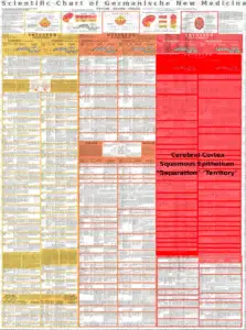
Cerebral cortex = New Brain = Outer Germ Layer = Ectoderm
Heart Attack – Diagnostic Chart
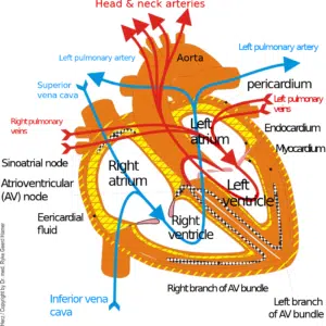
Coronary artery ulcers with severe angina.
Coronary arteries are (Heart Attack) pharyngeal duct derivatives and sensory supplied by the cortex.
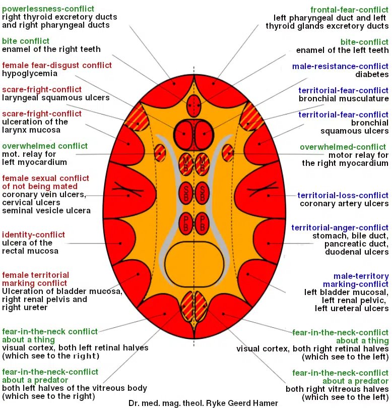
• In right-handed man: loss of territory conflict. Loss of the whole territory or the territory content. E.G.: Partner runs out of the territory.
• In a left-handed woman: Sexual conflict. Biological sexual frustration conflict of not being or having been mated is almost always accompanied by depression (even without hormonal “stalemate”).
• In schizophrenic constellation: With left-handed man and right-handed woman.
• In case of special hormonal situation, territorial conflict of male, right-handed woman in case of pill, postmenopause (so-called involution depression), castration, or masculinism. Territorial conflict with depression (resignation conflict) in weak, right-handed male in a hormonal stalemate.
“You’ve broken my heart!”
Right periinsular
Coronary artery ulcers with severe angina.
Swelling of the coronary artery intima, which is a squamous mucosa, in the area of the ulcers. Thereby coronary artery stenosis, which was mistakenly considered for the heart attack occurring after 2-6 weeks after CL. Male left heart attack = heart attack of the left heart.
Heart attack, absence, severe angina pectoris, cardiac arrest, and cardiac failure.
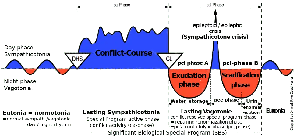
The so-called heart attack is the epileptic or epileptoid crisis that occurs 2-6 weeks after CL and proceeds more severely the longer and more intense the conflict has been. Symptoms: In a heart attack, there is maximal sympathicotonia. The entire conflict is experienced at a time-lapse pace during the coronary infarction.
The biological sense is to switch the vagotonia towards normotonia. If the sympathicotonic events are too weak, this switch cannot be successful. The “momentum” back to normality is missing. In conventional medicine, we used to give such patients sedatives and painkillers. Most of them died as a result. Today, we know that coronary pain during an infarction has its biological meaning and cannot be attenuated. It must not be attenuated.
Widening of the coronary arteries (due to ulceration) results in increased blood flow and efficiency.
Coronary arteries are pharyngeal duct derivatives and are supplied with cerebral sensibility.
CAUTION!!! Beware of the renal collecting tube syndrome: a large proportion of heart attack deaths may be due to the syndrome because the edema is significantly increased locally at the coronary and especially cerebrally in the territorial relay. The intra- and perifocal edema in the area of the HH is the reason for the heart attack and the possible cardiac arrest.
Therapy: High-dose intravenous administration of cortisone for the phase during or shortly after the epileptoid crisis, when the patient is usually at most significant mortal risk. No infusions. No vagotonic sedatives. CAUTION: the cortisone injection lasts only a few hours. After that, the danger may be as great again if no further injections follow; possibly switch to tablets.
The diagnostic chart dates from 2006, when Dr. Hamer prescribed cortisone for pulmonary embolism and myocardial infarction. The idea was to use the sympathicotonic effect of the cortisone to prevent the patient from going into vagotonia, thus intensifying the crisis and prolonging the healing phase. Then – according to the reasoning – the patient would more easily return to eutony after the crisis. A flat curve is easier to manage than a sharp one.
Dr. Hamer completely abandoned the use of cortisone in pulmonary embolisms and heart attacks. He discovered that cortisone does not have a purely sympathicotonic effect, like coffee. The cortisone acts as if a conflict relapse would have happened.
| Cookie | Duration | Description |
|---|---|---|
| cookielawinfo-checkbox-analytics | 11 months | This cookie is set by GDPR Cookie Consent plugin. The cookie is used to store the user consent for the cookies in the category "Analytics". |
| cookielawinfo-checkbox-functional | 11 months | The cookie is set by GDPR cookie consent to record the user consent for the cookies in the category "Functional". |
| cookielawinfo-checkbox-necessary | 11 months | This cookie is set by GDPR Cookie Consent plugin. The cookies is used to store the user consent for the cookies in the category "Necessary". |
| cookielawinfo-checkbox-others | 11 months | This cookie is set by GDPR Cookie Consent plugin. The cookie is used to store the user consent for the cookies in the category "Other. |
| cookielawinfo-checkbox-performance | 11 months | This cookie is set by GDPR Cookie Consent plugin. The cookie is used to store the user consent for the cookies in the category "Performance". |
| viewed_cookie_policy | 11 months | The cookie is set by the GDPR Cookie Consent plugin and is used to store whether or not user has consented to the use of cookies. It does not store any personal data. |
You’ll be informed by email when we post new articles and novelties. In every email there is a link to modify or cancel your subscription.