Not wanting to swallow a morsel, wanting to spit it out again.
for the right-handed person concerning partner
for the left-handed person concerning mother or child
Idiom:
–
Hamer Focus:
HH fronto parietal-basal left
Active phase:
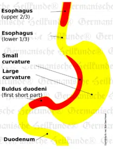
Ulcers in the upper esophageal region (upper 2/3). Since the squamous epithelium is very thick here, it takes 6 to 10 months before deep, visible ulcers are detectable gastroscopically. The esophagus is divided with respect to innervation (nervous supply), right and left are innervated crossed. Swallowing spasms.
Healing:
Swallowing stenoses. In this phase, the diagnosis is usually “papswallow stenosis” on X-ray. Now one would actually only have to wait for the end of the healing phase, since nothing more can actually happen. If the conflict lasts for a long time and/or is very intense, the swelling may cause swallowing difficulties. This can be remedied using a stomach tube through the nose for 2-3 months until the swelling has subsided.
Crisis:
Absence
Biological Sense:
Active phase
Ulcerative widening of the esophageal diameter.
We use cookies on our website to give you the most relevant experience by remembering your preferences and repeat visits. By clicking “Accept All”, you consent to the use of ALL the cookies. However, you may visit "Cookie Settings" to provide a controlled consent.
This website uses cookies to improve your experience while you navigate through the website. Out of these, the cookies that are categorized as necessary are stored on your browser as they are essential for the working of basic functionalities of the website. We also use third-party cookies that help us analyze and understand how you use this website. These cookies will be stored in your browser only with your consent. You also have the option to opt-out of these cookies. But opting out of some of these cookies may affect your browsing experience.
Necessary cookies are absolutely essential for the website to function properly. These cookies ensure basic functionalities and security features of the website, anonymously.
Cookie
Duration
Description
cookielawinfo-checkbox-analytics
11 months
This cookie is set by GDPR Cookie Consent plugin. The cookie is used to store the user consent for the cookies in the category "Analytics".
cookielawinfo-checkbox-functional
11 months
The cookie is set by GDPR cookie consent to record the user consent for the cookies in the category "Functional".
cookielawinfo-checkbox-necessary
11 months
This cookie is set by GDPR Cookie Consent plugin. The cookies is used to store the user consent for the cookies in the category "Necessary".
cookielawinfo-checkbox-others
11 months
This cookie is set by GDPR Cookie Consent plugin. The cookie is used to store the user consent for the cookies in the category "Other.
cookielawinfo-checkbox-performance
11 months
This cookie is set by GDPR Cookie Consent plugin. The cookie is used to store the user consent for the cookies in the category "Performance".
viewed_cookie_policy
11 months
The cookie is set by the GDPR Cookie Consent plugin and is used to store whether or not user has consented to the use of cookies. It does not store any personal data.
Functional cookies help to perform certain functionalities like sharing the content of the website on social media platforms, collect feedbacks, and other third-party features.
Performance cookies are used to understand and analyze the key performance indexes of the website which helps in delivering a better user experience for the visitors.
Analytical cookies are used to understand how visitors interact with the website. These cookies help provide information on metrics the number of visitors, bounce rate, traffic source, etc.
Advertisement cookies are used to provide visitors with relevant ads and marketing campaigns. These cookies track visitors across websites and collect information to provide customized ads.