Brainstem = Old Brain = Inner Germ Layer = Endoderm

Middle Ear Carcinoma left – Diagnostic Chart
The conflict dates back to ancient embryological times, when only one pharynx consisted of the middle ear and mouth.
–
In the brainstem (pons), left dorsal (backward) (nucleus of the so-called nervus stato-acusticus).
During cell proliferation, archaic hearing is quasi-enhanced. The archaic hearing organ receives the acoustic information. The flat-growing adeno-Ca of the absorptive grade grows only slightly in the middle ear and mastoid. The cells involved appear to be archaic auditory cells. In rare cases, after filling up the middle ear, the tumor may feign “regrowth,” i.e., imprint into the surrounding area (by impression).
Purulent otitis media (inflammation of the middle ear). Tuberculous-caseous necrotizing degradation of the increased cells by fungi or fungal bacteria (TBC) usually occurs with the perforation of the tympanic membrane (running ear). The healing has the sense to reduce the acoustic information back to normal because the acoustic morsel had been got rid of, and the conflict had been solved with it. The former so-called supposed bone conduction (tuning fork at the mastoid) was probably, for the most part, a function of the middle ear’s old intestinal cells together with the mastoid.
Centralization
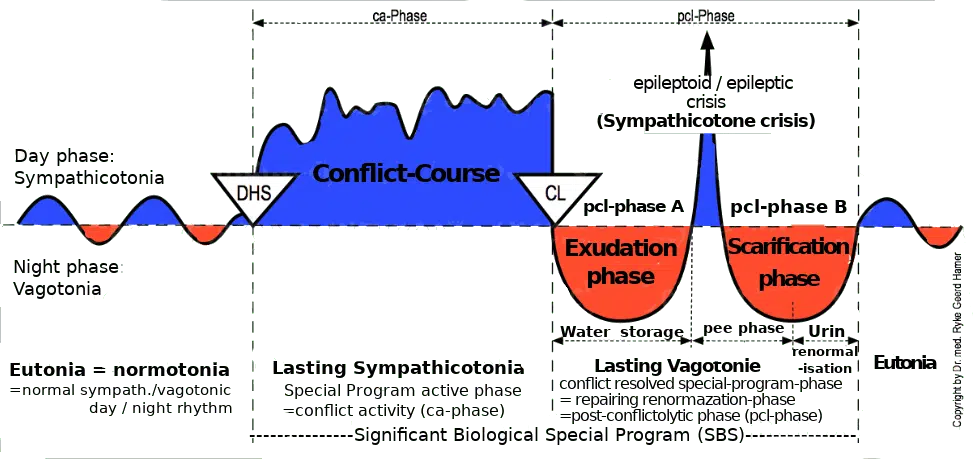
Active phase
During cell proliferation of the absorptive type, archaic hearing is quasi-enhanced by more acoustic information being received by the archaic auditory pathway.
–
| Cookie | Duration | Description |
|---|---|---|
| cookielawinfo-checkbox-analytics | 11 months | This cookie is set by GDPR Cookie Consent plugin. The cookie is used to store the user consent for the cookies in the category "Analytics". |
| cookielawinfo-checkbox-functional | 11 months | The cookie is set by GDPR cookie consent to record the user consent for the cookies in the category "Functional". |
| cookielawinfo-checkbox-necessary | 11 months | This cookie is set by GDPR Cookie Consent plugin. The cookies is used to store the user consent for the cookies in the category "Necessary". |
| cookielawinfo-checkbox-others | 11 months | This cookie is set by GDPR Cookie Consent plugin. The cookie is used to store the user consent for the cookies in the category "Other. |
| cookielawinfo-checkbox-performance | 11 months | This cookie is set by GDPR Cookie Consent plugin. The cookie is used to store the user consent for the cookies in the category "Performance". |
| viewed_cookie_policy | 11 months | The cookie is set by the GDPR Cookie Consent plugin and is used to store whether or not user has consented to the use of cookies. It does not store any personal data. |
You’ll be informed by email when we post new articles and novelties. In every email there is a link to modify or cancel your subscription.