“Modern” medicine had forgotten to examine the individual patient, not only his organs but also his psyche and brain. As a result, they have never been able to find a connection between psyche and organs, and mostly never between conflicts and organs.
With the tools of the trade, i.e., the knowledge of the five biological laws of Germanische Heilkunde®, and the knowledge of the respective typical symptoms of the course on the three levels, psyche-brain-organ, it is now possible for the first time in medicine to work causally and quasi reproducibly in a meaningful way. It is the classification according to the development history of embryology! Suppose we arrange all these different tumors, swellings, ulcers, or functional failures according to this development history or, according to their criteria, the different so-called germ leaves. In that case, everything suddenly arranges itself as if by itself. According to the so-called IRON RULE OF CANCER and the law of the two-phase nature of all diseases, when the conflict is resolved. This is the first systematic classification of all medicine.
The DHS (Conflict Shock) has become the linchpin of the entire Germanische Heilkunde®. It is beautiful that we can now really calculate and understand. We must mentally slip into the patient’s skin for this moment of the DHS and imagine how the overall situation was at the second of the DHS. The conflict content at the moment of the DHS determines both the localization of the Hamer Focus (HH) in the brain (so-called shooting target configuration) and cancer or cancer-equivalent disease, i.e., cancer-like disease at the organ. The biological conflicts are all archaic conflicts, apply analogously to humans and animals. Earlier, we considered the so-called “psychological conflicts,” better psychological problems, as the only essential conflicts. This was a mistake. Changes in the brain make only biological conflicts in humans and animals.
The law of biphasic of all diseases states that every disease has a conflict-active (ca-phase) and a conflict-resolved phase (pcl-phase) – provided that the conflict is resolved.
We know that all organs controlled by the alto-brain make cell proliferation (tumors) in the conflict-active phase from the ontogenetic system of tumors and cancer equivalents. All organs controlled by the cerebrum make cell reduction (necrosis, ulcers, holes, or loss of function) in the conflict-active phase.
This includes, among others, visual disturbances.

In the case of a fear-in-the-neck-conflict, which has its HH in the brain’s visual cortex, we have specific definition difficulties when we speak of cancer equivalent because the neurologists explain to us that the rods and cones of the retina basically still belong to the brain. In any case, it is certain that on the psychological and cerebral level, all five laws of Germanische Heilkunde® are precisely fulfilled.
As is known, the optic nerve fibers partially cross. The left visual cortex receives all rays coming from the left (and falling on the right retinal halves of both eyes). The right visual cortex receives all rays coming from the right (and falling on the left retinal halves of both eyes). However, the fibers from the fovea centralis belong to the lateral half and therefore guide the images predominantly to the opposite visual cortex.

To understand the psychological side of biological conflicts, one must trace them back together to organ manifestation development historically. All designations of these biological conflicts are so selected that they can have validity at the same time for the mammal (real) and humans in the possibly transferred sense.
Fear-in-the-neck-conflict means a danger that one cannot face, which continually threatens or lurks from behind and cannot shake off.
In the conflict-active phase, HH in the right or left visual cortex occipital, for the retinal halves. It results in progressive loss of the visual ability of a particular retinal relay.
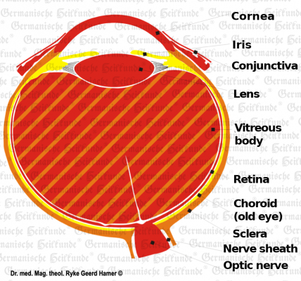
In the healing phase, the obligatory edema is formed in the HH of the visual cortex and between the sclera and the retina. The healing edema is formed, which leads to the so-called retinal detachment. Although this is a good healing symptom, and even if the conflict does not last too long, it is reversible, i.e., it also recedes on its own. Still, initially, there is a dramatic deterioration of vision precisely due to this retinal detachment.
Myopia results in lateral retinal detachments with several recurrences, resulting in optical lengthening of the eyeball because the retinal detachment is fixed by occlusion between the retina and the sclera.
In the case of dorsal retinal detachment, with several recurrences and consequent maintenance between the retina and the sclera, which results in optical shortening of the eyeball, farsightedness results.
If the retinal detachment occurs at the point of sharpest vision, it is called macular degeneration.
If both visual cortices are affected, i.e., two HH are active in the right and left visual cortex (corresponding to two conflicts of the fear in the neck). The pat. is in a so-called schizophrenic constellation and has a persecution delusion, which is not as crazy as we used to think but represents an attempt to get rid of the fear in the neck, i.e., to solve the conflict. The pat. consistently avoids all occasions, however minor because of his “delusion,” which we just did not understand until now.
Fear-in-the-neck-conflict or with remarkable aspect, affecting the paramedian part of the visual cortex, means that the anxiety is felt behind the eye, as the consciousness’s orientation center.
In the conflict-active phase, a partial clouding of the vitreous body takes place. The biological sense is that with the eyes of the so-called prey animals, which usually look to the side, the danger from behind is quasi-covered or obscured. Still, the view to the front on the escape route remains free so that the prey animal finds its escape route to the front sure-footedly, without always looking back in panic after the predator. A “fogging” of the backward vision occurs, a partial clouding of the vitreous body, so-called “glaucoma.” Therefore, only a part of the vitreous body is clouded (blinker phenomenon). The predators can afford to look forward with both eyes because they have to be afraid of another predator to a much lesser extent.
In the healing phase, the vitreous opacity also regresses, with vitreous edema formation, a so-called glaucoma formation, and increased pressure inside the eye. Often the edema pushes back through the hole of the entrance of the optic nerve. Laser treatment is not allowed in the ca-phase or healing phase. This would irreparably destroy the vitreous body.
Example: A patient experienced an assault in which a man tried to rape her in the dark on her way home from the subway. She immediately got into several conflicts. When she tried to run away, and the man came from behind, she suffered a fear-in-the-neck conflict from the robber (the rapist). The patient got recurrences for years. The conflict remained active for years because she always had to take the same subway to work and always had the same way home. Even in winter, when it got dark early, she saw a rapist lurking behind every bush. She had no idea that it was this conflict track that clouded both her vitreous (glaucoma).
The eye lens has nothing to do with the visual cortex but conflictively corresponds to a strong visual separation conflict (when one loses sight of someone). We see necrosis of the lens in the conflict-active phase. The Hamer Focus here is in the sensory cortex center. The Hamer’s focus here is in the sensory cortex center. The trigeminal 1st branch (ophtalmicus) is located in the cerebrum laterally, and temporally. The biological sense is that you can follow someone walking away from you, strange as it may sound, further with your eyes with a necrotized lens than with an intact lens. This gives the person even better distance vision. And in the healing phase, when the individual is lost from sight (human or animal) is back, the lens temporarily becomes cloudy (as a sign of healing), which we then call a cataract. If this conflict runs only once, the lens clears up again. However, if there are several recurrences, the clouding increases more and more. However, this is not understood in current medicine, especially since the patient can become blind in the process. If one waited accordingly, the lens would spontaneously clear again, provided the conflict remains solved. So-called glaucoma, the increase in the intraocular pressure, can occur both in the healing phase of the vitreous opacity and in the healing phase of the lens necrosis. But glaucoma is always a sign of the healing phase.
The underlying conflict is a strong visual separation conflict (losing someone from sight). The corneal ulcers are replenished in the healing phase with temporary corneal opacity: keratitis.
A conflict of separation also accompanies the epithelial ulcer of the eyelids and conjunctiva. Ulcers of the eyelids and conjunctiva form in the conflict-active phase: scaling. In the healing phase, reddening of the eyelids (blepharitis) and reddening of the conjunctiva (conjunctivitis) occur.

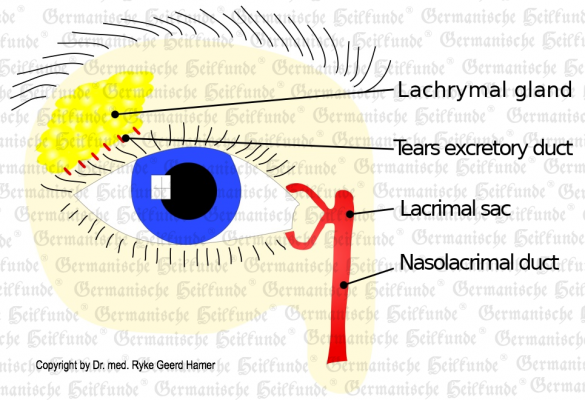
In lacrimal duct excretory duct ulcer-Ca, we always find a conflict of wanting to be seen or not wanting to be seen with a HH frontomedio-lateral-basal. In the conflict-active phase, ulcers form in the ectodermal excretory ducts of the lacrimal glands. In the healing phase, the mucosa in the excretory ducts swells, causing congestion and severe swelling of the entire lacrimal gland. This seems like a tumor of the lacrimal gland, which is not.
In contrast, the lacrimal gland-Ca (acinar part) has its relay in the brainstem (pons), thus belongs to the alto-brain (inner germ layer) and therefore makes – like all alto-brain-controlled organs – cell proliferation (tumors), in the conflict-active phase. Here, the conflict of “not being able to grasp the lump because one is not seen and thus passed over” is the basis.
Example: A painter does not find a gallery owner, his pictures are not seen, he cannot sell anything and thus does not get hold of the lump (sales proceeds).
I.e., biologically: With the animal, it goes with a morsel, which it does not get to grasp, really around a piece of food. With humans (patient), it can be just as well the sales proceeds (pictures), with which it must finally procure its food. But if nobody is interested in his pictures, he also gets no money (morsel) and can buy no food.
There are also acini in the lacrimal glands that produce the tear fluid. In extreme cases, these degenerate into compact tumors that can bulge the eyelid outward. In the healing phase, both encapsulation and caseation with fetid discharge are possible (cold abscess, cold abscess), and purulent tears (caused by fungi and fungal bacteria). In the case of total caseation (recurrences) of the glandular cells, the result is cystic duct disease of the lacrimal glands, i.e., drying up the lacrimal fluid.
Copyright © 2024 by GHk Online Service, LLC
| Cookie | Duration | Description |
|---|---|---|
| cookielawinfo-checkbox-analytics | 11 months | This cookie is set by GDPR Cookie Consent plugin. The cookie is used to store the user consent for the cookies in the category "Analytics". |
| cookielawinfo-checkbox-functional | 11 months | The cookie is set by GDPR cookie consent to record the user consent for the cookies in the category "Functional". |
| cookielawinfo-checkbox-necessary | 11 months | This cookie is set by GDPR Cookie Consent plugin. The cookies is used to store the user consent for the cookies in the category "Necessary". |
| cookielawinfo-checkbox-others | 11 months | This cookie is set by GDPR Cookie Consent plugin. The cookie is used to store the user consent for the cookies in the category "Other. |
| cookielawinfo-checkbox-performance | 11 months | This cookie is set by GDPR Cookie Consent plugin. The cookie is used to store the user consent for the cookies in the category "Performance". |
| viewed_cookie_policy | 11 months | The cookie is set by the GDPR Cookie Consent plugin and is used to store whether or not user has consented to the use of cookies. It does not store any personal data. |
You’ll be informed by email when we post new articles and novelties. In every email there is a link to modify or cancel your subscription.