
Cerebellum = Old Brain = Middle Germ Layer = Mesoderm
Pleura Carcinoma right
Conflict of attack against the inner thoracic cavity.
Example: “You have a lung tumor, you need surgery.” Or: Surgeon: “We have to pry open your chest to get to it.” Both a real attack suffered and an attack threatened or imagined are possible.
–
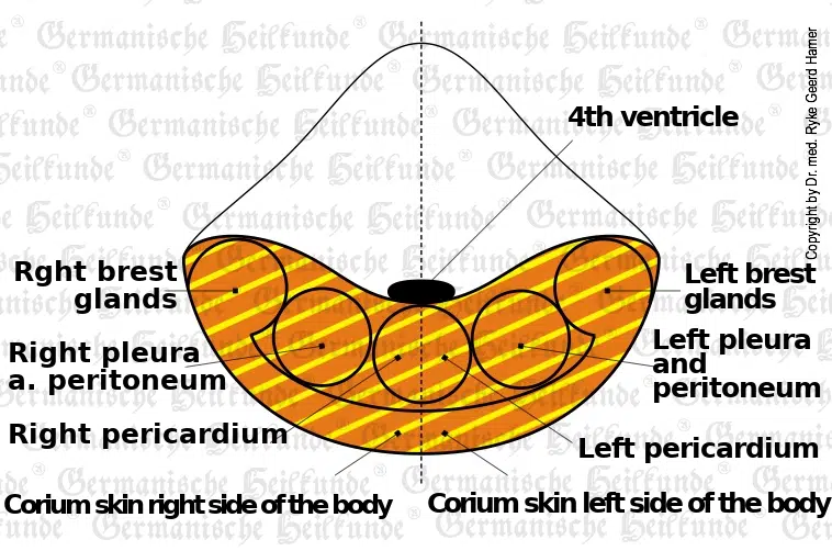
HH in the mid-lateral cerebellar area on the left, pleura, and peritoneum are in the same location in the cerebellum and are difficult to distinguish.
The compact mesotheliomas of the pleura can grow areal or make single, compact, large tumors, depending on the type of attack felt. For example, suppose the patient sees an X-ray of the lung during DHS, and there is a tumor on the left. In that case, he suffers the mesothelioma and subsequent pleural effusion on the left. However, the tumor (e.g., bronchial-Ca) was actually located on the right.
Pleural effusion is a sign of healing. The respiratory problems occur only in large pleural effusion, partly due to the effusion itself, partly due to the cerebral edema. The pleural effusion is noticed all the more dramatic when there is an active refugee conflict at the same time (syndrome). Most pleural effusions, both exudative and transudative, would have little or no clinical manifestation without the syndrome. This is especially true in the case of transudative pleural effusion with the syndrome, which should correctly be called “gout” (see under healing phase of bone osteolysis or leukemia). After expired TBC, we often find the usual calcium deposits.
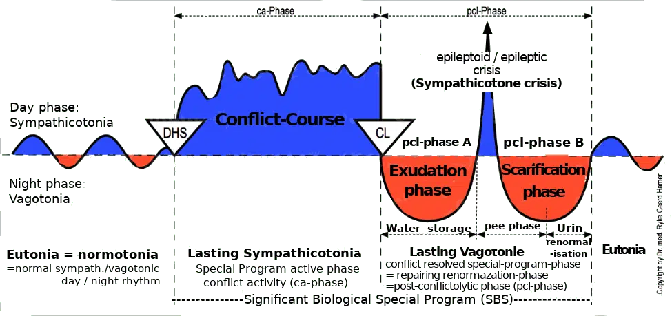
Centralization
Active phase
Thickening of the pleura to prevent an expected stab or punch; also mental: “You have a lung tumor!”
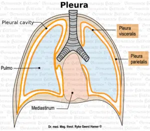
We distinguish between a parietal pleura (lining of the thoracic cavity, so-called pleura) and a visceral pleura (covering the lungs).
| Cookie | Duration | Description |
|---|---|---|
| cookielawinfo-checkbox-analytics | 11 months | This cookie is set by GDPR Cookie Consent plugin. The cookie is used to store the user consent for the cookies in the category "Analytics". |
| cookielawinfo-checkbox-functional | 11 months | The cookie is set by GDPR cookie consent to record the user consent for the cookies in the category "Functional". |
| cookielawinfo-checkbox-necessary | 11 months | This cookie is set by GDPR Cookie Consent plugin. The cookies is used to store the user consent for the cookies in the category "Necessary". |
| cookielawinfo-checkbox-others | 11 months | This cookie is set by GDPR Cookie Consent plugin. The cookie is used to store the user consent for the cookies in the category "Other. |
| cookielawinfo-checkbox-performance | 11 months | This cookie is set by GDPR Cookie Consent plugin. The cookie is used to store the user consent for the cookies in the category "Performance". |
| viewed_cookie_policy | 11 months | The cookie is set by the GDPR Cookie Consent plugin and is used to store whether or not user has consented to the use of cookies. It does not store any personal data. |
You’ll be informed by email when we post new articles and novelties. In every email there is a link to modify or cancel your subscription.